














Anesthesia Reveals Unexpected Levels of Brain Activity and Consciousness
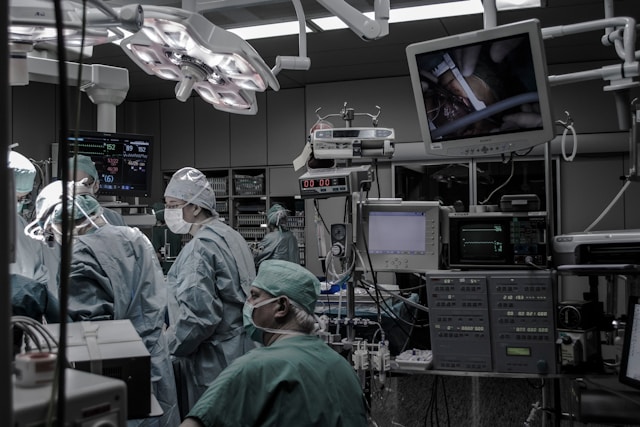
Seeing the unseen: neuroimaging uncovers language processing while patients are unconscious
When a surgeon turns the knob on a ventilator and a patient slips into a state that feels, by definition, “no‑thing‑going‑on,” the operating room lights still flash, monitors still beep, and a team of clinicians still watches. Yet a series of recent studies using functional magnetic resonance imaging (fMRI) and magnetoencephalography (MEG) have shown that the brain does not simply shut off. In a landmark experiment at Baylor College of Medicine, volunteers listened to spoken stories while a standard cocktail of propofol and sevoflurane kept them clinically unresponsive. The imaging data revealed that auditory cortex continued to track the rhythm of speech, that higher‑order temporal regions decoded word meaning, and that prefrontal areas prepared predictions about upcoming sentences—processes that, under normal waking conditions, support comprehension and memory formation (ScienceDaily, 2026). The researchers describe the phenomenon as “brain activity under anesthesia” that is far richer than the flat silence once assumed.
How the experiments measured lingering cognition
To capture fast, hierarchical processing the investigators paired high‑resolution fMRI with a “naturalistic” stimulus: a 10‑minute narrative read aloud. Participants first underwent a baseline scan while awake, then received a dose calibrated to achieve a bispectral index (BIS) score below 40, the clinical threshold for loss of consciousness. While the anesthetic level remained steady, the story continued. Voxel‑wise analyses showed that primary auditory cortex retained a reliable hemodynamic response to the acoustic envelope of the speech. More strikingly, multivariate pattern classifiers could still distinguish between semantically related and unrelated word clusters in the superior temporal gyrus, indicating that the brain preserved a level of semantic discrimination (ScienceDaily, 2026).
MEG recordings from a separate cohort added temporal precision. Event‑related fields demonstrated that the brain’s “N400” component—a marker of semantic integration—persisted, albeit with reduced amplitude, during the anesthetized state. This suggests that the neural machinery that binds words to meaning does not vanish entirely when consciousness is pharmacologically suppressed (ScienceDaily, 2026). The authors stress that the observed activity is not merely residual noise; the patterns tracked the narrative’s structure and predicted upcoming linguistic units, a hallmark of active processing.
What the data reveal about residual cognition
The finding that language networks stay online raises the question “is there brain activity under anesthesia?” The answer, according to the new work, is unequivocally yes—though the activity is altered in amplitude and synchrony. Parallel animal work has shown that certain cortical cell types become highly synchronized when exposed to anesthetic agents. In mice, layer‑5 pyramidal neurons across the cortex fire in a coordinated, aperiodic rhythm that is absent in other cell classes (Bharioke et al., 2022). This synchrony may provide a scaffold that preserves the ability of higher‑order circuits to exchange information, even as overall firing rates drop.
Human imaging studies corroborate a shift in the brain’s temporal‑spatial organization. During anesthesia, the long‑range temporal correlations that normally link distant regions weaken, producing a more fragmented pattern of activity (Zhang et al., 2018). The same work points to a “temporospatial theory of consciousness” that ties the richness of conscious experience to nested, hierarchical dynamics. The Baylor findings suggest that while the global nesting collapses, pockets of organized processing survive, perhaps because they are anchored in the synchronized layer‑5 activity observed in rodents.
Neurochemical investigations add another layer. Dopamine, a neurotransmitter linked to wakefulness, appears to modulate the depth of anesthetic suppression. In rodent models, boosting dopaminergic signaling hastens emergence from anesthesia, implying that the dopaminergic system helps sustain the “ignition” of cortical networks (Wang et al., 2023). By contrast, glutamatergic neurons—responsible for most excitatory signaling—show reduced firing under anesthetic drugs, a pattern that mirrors the transition from wake to sleep (Luo et al., 2025). The coexistence of suppressed excitation and preserved synchrony may explain why some cognitive operations persist while others fade.
Rethinking consciousness: where the findings sit among leading theories
Philosophers and neuroscientists have long debated whether consciousness is a binary switch or a graded phenomenon. The classic view, rooted in clinical practice, treats loss of consciousness as an all‑or‑none event: the patient is either responsive or not. The new neuroimaging data challenge that binary framing. If language processing can continue without overt awareness, consciousness may be better understood as a spectrum of functional capacities, each with its own neural signature.
One influential account, the Global Workspace Theory (GWT), proposes that conscious experience arises when information becomes globally available across the brain’s frontoparietal network (Dehaene, 2011). The anesthetized brain shows reduced global integration, yet retains localized processing in auditory and temporal cortices. This pattern fits a “partial workspace” scenario: information circulates within a limited circuit but fails to reach the full broadcast that characterizes waking consciousness.
Integrated Information Theory (IIT) takes a different angle, quantifying consciousness in terms of the system’s ability to generate irreducible cause–effect structures (Seth, 2022). The observed drop in long‑range temporal correlations would lower the system’s integrated information (Φ), consistent with a reduced conscious level. However, the persistence of structured activity in language areas suggests that Φ does not fall to zero; instead, it may settle at an intermediate value reflecting a “sub‑conscious” regime.
Higher‑order theories argue that a mental state becomes conscious only when it is the object of another, higher‑order representation (Block, 1995). Under anesthesia, the brain appears unable to generate those meta‑representations, even though first‑order sensory processing continues. This aligns with the clinical observation that patients cannot later recall the story they heard while anesthetized—a hallmark of absent higher‑order tagging.
Even the philosophical “what is it like?” question posed by Nagel (1974) finds new context. If a patient’s brain processes language without the accompanying subjective feeling, the “what it is like” component may be missing, suggesting that consciousness hinges on more than raw information flow. The anesthetic data therefore sharpen the distinction between neural computation and phenomenology.
Clinical ripples: anesthesia monitoring and patient safety
Surgeons and anesthesiologists have long relied on behavioral signs—movement, eye opening, hemodynamic changes—to gauge depth of anesthesia. The discovery that meaningful brain activity persists raises practical concerns. Could a patient be “awake” enough to experience pain or form memories without showing overt signs? While the studies to date have not demonstrated explicit awareness under the drug regimens used, they do suggest that the brain’s capacity to encode information remains partially intact.
One immediate implication is the potential refinement of intra‑operative monitoring. Electroencephalography (EEG) based indices such as BIS already track the overall suppression of cortical activity, but they may miss localized processing. Incorporating measures of auditory evoked potentials or tracking the integrity of the N400 component could provide a more nuanced picture of the patient’s neural state. Such metrics would answer the lay query “what happens to brain activity under anesthesia?” with a data‑driven response rather than a blanket “nothing happens.”
Another area of interest is the choice of anesthetic agents for neurosurgical procedures. Certain drugs, like propofol, preferentially enhance GABA‑mediated inhibition, while others, such as ketamine, act on NMDA receptors and can produce dissociative states where higher‑order processing is more preserved (Hu, 2024). When operating on the brain itself, surgeons sometimes use “awake” techniques with minimal sedation to allow intra‑operative mapping; the new findings suggest that even low‑dose regimens might leave language circuits partially active, a factor that could be harnessed or mitigated depending on the surgical goal.
From a patient‑communication standpoint, the question “do you have brain activity under anesthesia?” can now be answered with nuance: yes, but the activity does not equate to conscious experience. Explaining this distinction may alleviate postoperative anxiety about “intra‑operative awareness,” a rare but feared complication. Moreover, understanding the residual processing could inform postoperative care, as lingering neural activity might influence how memories of the surgical event are consolidated—or not.
Open questions and next steps
Several avenues remain underexplored. First, the current studies used relatively simple auditory narratives; it is unknown whether more complex cognitive tasks—such as problem solving or emotional appraisal—survive anesthesia to a similar degree. Second, the animal work points to layer‑specific synchrony as a possible substrate for residual processing, yet direct translation to human cortical layers is technically challenging. Advances in high‑field MRI and laminar electrophysiology may soon bridge that gap.
Third, the role of neuromodulatory systems warrants deeper investigation. Dopamine appears to facilitate emergence, but whether manipulating dopaminergic tone during surgery could preserve beneficial processing without risking awareness is an open clinical question (Wang et al., 2023). Likewise, the balance between glutamatergic suppression and GABAergic enhancement shapes the anesthetic landscape; fine‑tuning this balance could allow surgeons to target specific networks while keeping others quiet (Luo et al., 2025).
Finally, the theoretical implications call for interdisciplinary dialogue. The data provide empirical fodder for debates that have traditionally unfolded in philosophy departments. By mapping the precise neural signatures that survive anesthetic suppression, researchers can test predictions derived from GWT, IIT, and higher‑order theories in a controlled, reversible setting. Such experiments could move the field beyond speculative argument to a more empirical grounding of consciousness research.
In practice, the take‑home message for clinicians and patients alike is that “brain activity under general anesthesia” is a richer phrase than “nothing happening.” The brain continues to parse language, to anticipate what comes next, and to maintain a baseline of organized firing even when the patient cannot report any experience. Recognizing this nuance reshapes how we think about unconsciousness, informs safer anesthetic practices, and opens a window onto the neural architecture of consciousness itself.
References
- Bharioke. (2022). General anesthesia globally synchronizes activity selectively in layer 5 cortical pyramidal neurons. Neuron. https://doi.org/10.1016/j.neuron.2022.03.032
- Zhang. (2018). Breakdown in the temporal and spatial organization of spontaneous brain activity during general anesthesia. Human Brain Mapping. https://doi.org/10.1002/hbm.23984
- Wang. (2023). Dopaminergic System in Promoting Recovery from General Anesthesia. Brain Science. https://doi.org/10.3390/brainsci13040538
- Hu. (2024). Neural Network Mechanisms Underlying General Anesthesia: Cortical and Subcortical Nuclei. Neuroscience Bulletin. https://doi.org/10.1007/s12264-024-01286-z
- Luo. (2025). The regulation of glutamatergic nervous system in sleep-wake states and general anesthesia.. Brain Research Bulletin. https://doi.org/10.1016/j.brainresbull.2025.111220
- Takla. (2021). General anaesthesia in end-of-life care: extending the indications for anaesthesia beyond surgery.. Anaesthesia. https://doi.org/10.1111/anae.15459
- (2015). Medical Renaissance. Journal of Investigative Surgery. https://doi.org/10.3109/08941939.2015.1054747
- (2012). Asepsis and bacteriology: a realignment of surgery and laboratory science. Medical History. https://doi.org/10.1017/mdh.2012.22
- (2017). Two Japanese Pioneers in Anesthesiology: Seishū Hanaoka and Gendai Kamada. Journal of Anesthesia History. https://doi.org/10.1016/j.janh.2016.12.002
- (1937). The Stages and Signs of General Anaesthesia. British Medical Journal. https://doi.org/10.1136/bmj.2.3996.274
- Chalmers. (1996). The Conscious Mind: In Search of a Fundamental Theory. Oxford University Press.
- Nagel. (1974). What Is It Like to Be a Bat?. The Philosophical Review. https://doi.org/10.2307/2183914
- Dehaene. (2011). Experimental and Theoretical Approaches to Conscious Processing. Neuron. https://doi.org/10.1016/j.neuron.2011.03.018
- Seth. (2022). Theories of consciousness. Nature Reviews Neuroscience. https://doi.org/10.1038/s41583-022-00587-4
- Block. (1995). On a Confusion about a Function of Consciousness. Behavioral and Brain Sciences. https://doi.org/10.1017/S0140525X00038188
Paper data via Semantic Scholar.