


















Mini Brains in a Dish: The Future of Alzheimer’s Drug Testing
INTRODUCTION
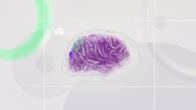
When a loved one receives an Alzheimer’s diagnosis, the uncertainty can feel like staring into a fogged‑up mirror—every day brings new questions about what treatments might actually help. Imagine if doctors could look at a tiny, living replica of that person’s brain and see, in real time, which medication eases the fog and which does nothing at all. That vision is moving from science‑fiction toward reality thanks to a breakthrough in Alzheimer's organoid drug testing. In a recent study, researchers grew miniature brain models from patients’ own cells and observed drug responses that mirrored what happens in the full organ. The implication? A future where treatment plans are tailored to the individual, and the long, costly road of drug development could be shortened dramatically. Below we unpack how these “mini brains” are made, why they matter beyond the lab, and what this could mean for families navigating the challenges of Alzheimer’s.
How Alzheimer's organoid drug testing is reshaping research
Cerebral organoids are three‑dimensional clusters of neural tissue that develop from pluripotent stem cells—cells capable of becoming any cell type in the body. By coaxing these stem cells with specific chemical cues, scientists can guide them to self‑organize into structures that resemble early‑stage human brain regions, complete with neurons and supporting glial cells (Cerebral organoids model, 2013). Over the past decade, advances in culture techniques have allowed organoids to mature for months, reaching developmental stages comparable to the early post‑natal brain (Long-term maturation of, 2021). This prolonged maturation is crucial for modeling neurodegenerative diseases, which unfold over years in patients.
Traditionally, Alzheimer’s research has relied on rodent models or two‑dimensional cell cultures. While useful, these systems fall short in several ways. Rodent brains differ markedly from human brains in cellular composition, gene expression, and the way they accumulate amyloid plaques and tau tangles—the hallmark protein aggregates of Alzheimer’s (Alzheimer's disease, 2009). Two‑dimensional cultures lack the spatial architecture that influences cell‑cell communication and drug diffusion. In contrast, patient‑derived organoids preserve the individual’s unique genetic background, including risk alleles such as APOE ε4, and recapitulate the three‑dimensional microenvironment of the human cortex (The use of, 2017). This makes them a more faithful platform for testing therapeutic agents.
In the Johns Hopkins study, skin fibroblasts from several Alzheimer’s patients were reprogrammed into induced pluripotent stem cells (iPSCs) and then differentiated into cortical organoids. After roughly four to six months of growth, the organoids displayed classic Alzheimer’s pathology: extracellular amyloid‑β deposits and intracellular phosphorylated tau aggregates. The researchers then exposed the organoids to a serotonergic antidepressant that has shown modest cognitive benefits in early clinical trials. Strikingly, organoids derived from different patients exhibited divergent biochemical responses—some showed a reduction in amyloid‑β secretion, while others showed no change or even a paradoxical increase. These divergent patterns echoed the variable clinical outcomes observed in patients receiving the same drug, suggesting that organoid responses can predict individual therapeutic efficacy (The use of, 2017).
Beyond the drug‑specific findings, the study demonstrated a pipeline that could be scaled for high‑throughput screening. By standardizing the differentiation protocol, researchers can generate thousands of near‑identical organoids, each representing a distinct patient genotype (Being Patient). This scalability opens the door to “personalized phenotypic screens,” where a library of candidate compounds is tested across a panel of patient‑specific organoids to identify those that consistently normalize disease markers. Such an approach aligns with modern phenotypic drug discovery, which emphasizes observing functional outcomes in complex cellular systems rather than targeting a single molecular interaction (Modern phenotypic drug, 2012).
Why does this matter for drug development economics? Traditional Alzheimer’s drug pipelines are notoriously expensive and slow. The average cost to bring a new molecular entity to market exceeds $1.8 billion, with a high attrition rate during late‑stage clinical trials (How to improve, 2010). A major cause of failure is the mismatch between animal model predictions and human outcomes. By integrating organoid‑based screening early in the pipeline, pharmaceutical companies could deprioritize compounds that lack efficacy in human‑like tissue, thereby conserving resources and accelerating the arrival of promising therapies to patients (Classical vs reverse, 2001).
Importantly, organoid platforms also enable the study of disease mechanisms that are difficult to capture in animals. For example, the interplay between amyloid pathology and neuroinflammation can be examined by co‑culturing microglia‑like cells with organoids, revealing how certain drugs modulate immune responses in a patient‑specific context (Cerebral organoids as, 2020). Such mechanistic insights can guide the rational design of combination therapies that target multiple disease pathways simultaneously.
Why this breakthrough matters to everyday lives
For families confronting Alzheimer’s, the promise of personalized treatment is more than a scientific curiosity—it offers tangible hope. Currently, clinicians prescribe the same handful of FDA‑approved drugs (e.g., cholinesterase inhibitors, NMDA receptor antagonists) to all patients, despite well‑documented variability in how individuals respond (Alzheimer's disease, 2009). This one‑size‑fits‑all approach can lead to months of trial‑and‑error, during which cognitive decline may continue unchecked.
With Alzheimer’s organoid drug testing, a neurologist could, in principle, obtain a small skin biopsy from a patient, generate a personalized organoid, and test a panel of approved and experimental compounds within weeks. The organoid’s biochemical readouts—levels of amyloid‑β, tau phosphorylation, synaptic protein expression—would inform the clinician which medication is most likely to stabilize or improve the patient’s cognition. This could shorten the “guesswork” period, allowing patients to spend more time with clearer minds and less time coping with side‑effects from ineffective drugs.
Beyond individual treatment, organoid data can inform public health strategies. If certain genetic sub‑groups consistently respond to a specific class of drugs, screening programs could prioritize those therapies for at‑risk populations, optimizing resource allocation in healthcare systems strained by the growing prevalence of dementia (Survival in dementia, 2013). Moreover, faster drug validation reduces the time it takes for novel therapies to reach market, potentially lowering costs for insurers and patients alike.
From a societal perspective, accelerating effective treatments could also alleviate the enormous caregiver burden associated with Alzheimer’s. Families often shoulder 15–20 hours of care per week, leading to emotional stress, reduced work productivity, and financial strain (Dementia in Down, 2019). By delivering therapies that meaningfully slow disease progression, organoid‑guided precision medicine could preserve patient independence longer, granting families more quality time together and reducing the long‑term economic impact of the disease.
Practical takeaways for patients, caregivers, and clinicians
1. Ask about clinical trials that incorporate organoid testing. Many academic medical centers are now enrolling participants in studies where a patient‑derived organoid is used to screen investigational drugs. Inquiring with your neurologist can provide access to cutting‑edge personalized therapy options.
2. Advocate for genetic counseling. Since organoid responses are influenced by a patient’s genetic makeup, understanding one’s APOE status and other risk alleles can help interpret organoid‑based recommendations and guide lifestyle interventions that may complement pharmacologic treatment.
3. Support research participation. Donating a small skin sample for iPSC generation is minimally invasive and contributes to a growing biobank of diverse organoids. A broader representation of ethnic and age groups improves the predictive power of organoid drug screens for the entire population.
4. Stay informed about emerging biomarkers. As organoid platforms mature, they are being paired with fluid biomarkers (e.g., plasma amyloid‑β, neurofilament light) to create composite predictive models. Keeping abreast of these developments can help you discuss comprehensive monitoring plans with your care team.
Closing thoughts
Imagine a future where the fog of Alzheimer’s can be lifted not by trial and error, but by a tiny, patient‑specific brain in a dish that tells us exactly which medication will work. The early successes of Alzheimer’s organoid drug testing suggest that such a future is within reach. While the technology is still moving from the lab bench toward the clinic, its potential to personalize therapy, streamline drug development, and ultimately give families more time with their loved ones is profound. As research continues to refine these mini brains, the hope is that they will become a standard tool in the neurologist’s arsenal—turning the once‑opaque landscape of Alzheimer’s treatment into a clearer, more navigable path for everyone affected.
References
- (2013). Cerebral organoids model human brain development and microcephaly. Nature. https://doi.org/10.1038/nature12517
- (2021). Long-term maturation of human cortical organoids matches key early postnatal transitions. Nat Neurosci. https://doi.org/10.1038/s41593-021-00802-y
- (2017). The use of brain organoids to investigate neural development and disease. Nature Reviews. Neuroscience. https://doi.org/10.1038/nrn.2017.107
- (2007). Neuroscience.. W. H. Freeman.
- (2020). Cerebral organoids as tools to identify the developmental roots of autism. Molecular Autism. https://doi.org/10.1186/s13229-020-00360-3
- (2016). Lewy Body Dementias: Dementia With Lewy Bodies and Parkinson Disease Dementia. Continuum. https://doi.org/10.1212/CON.0000000000000309
- (2019). Dementia in Down syndrome: unique insights for Alzheimer disease research. Nat Rev Neurol. https://doi.org/10.1038/s41582-018-0132-6
- (2013). Survival in dementia and predictors of mortality: a review. International Journal of Geriatric Psychiatry. https://doi.org/10.1002/gps.3946
- (2009). Alzheimer's disease. BMJ. https://doi.org/10.1136/bmj.b158
- Zheng. (2024). Predictors for survival in patients with Alzheimer's disease: a large comprehensive meta-analysis. Translational Psychiatry. https://doi.org/10.1038/s41398-024-02897-w
- (2010). How to improve R&D productivity: the pharmaceutical industry's grand challenge. Nature Reviews. Drug Discovery. https://doi.org/10.1038/nrd3078
- (2009). Current Model for Financing Drug Development: From Concept Through Approval. Institute of Medicine (US), Forum on Drug Discovery, Development, and Translation, National Academies Press, Washington (DC).
- (2011). Drug discovery: lessons from evolution. British Journal of Clinical Pharmacology. https://doi.org/10.1111/j.1365-2125.2010.03854.x
- (2001). Classical vs reverse pharmacology in drug discovery. BJU International. https://doi.org/10.1111/j.1464-410X.2001.00112.x
- (2012). Modern phenotypic drug discovery is a viable, neoclassic pharma strategy. Journal of Medicinal Chemistry. https://doi.org/10.1021/jm201649s